
Electron Microscopy
Access:
Seeing biology at molecular resolution—from purified complexes to whole cells
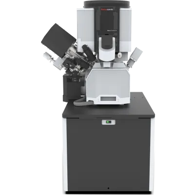
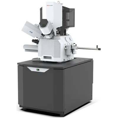
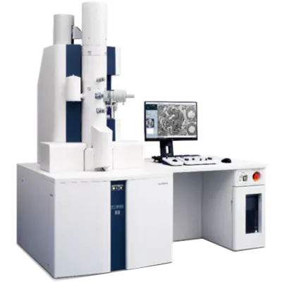
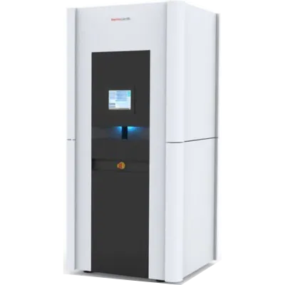
NYSBC operates one of the world’s most comprehensive platforms for cryo-electron microscopy. It encompasses four main equipment ecosystems: transmission electron microscopes optimized for single-particle imaging and cellular tomography; cryo-focused ion beam systems for in situ sample preparation; integrated light microscopy for correlative workflows; and a complete set of sample preparation and supporting technologies. Together, these capabilities enable researchers to visualize molecular structure across the full range of biological complexity—from purified assemblies at near-atomic resolution to intact cellular environments in their native state. With all of these tools under one management structure, expertise and data workflows are shared. Users can thus access complementary capabilities seamlessly, moving from initial sample screening to structure determination and through to manuscript-ready analysis.
The facility’s approach integrates cutting-edge instrumentation with the development of methods that reduce barriers to use. Whether scientists are imaging a well-characterized complex or exploring new territory, NYSBC’s staff work alongside them to optimize sample preparation, data collection, and analysis. Member institutions access the facility directly; national researchers apply through NCCAT or NCITU.
Single Particle Cryo-Electron Microscopy
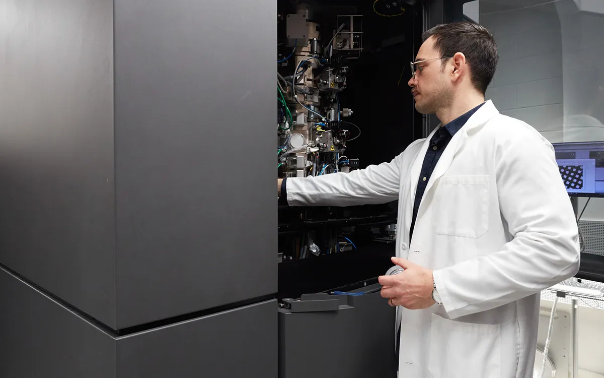
Cryo-Electron Tomography
Cryo-FIB (Focused Ion Beam)

Correlative Light and Electron Microscopy (CLEM)

















Equipment
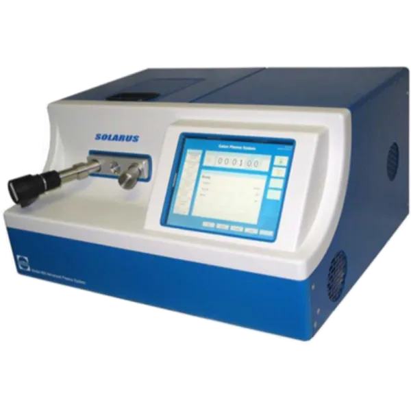
Gatan Solarus
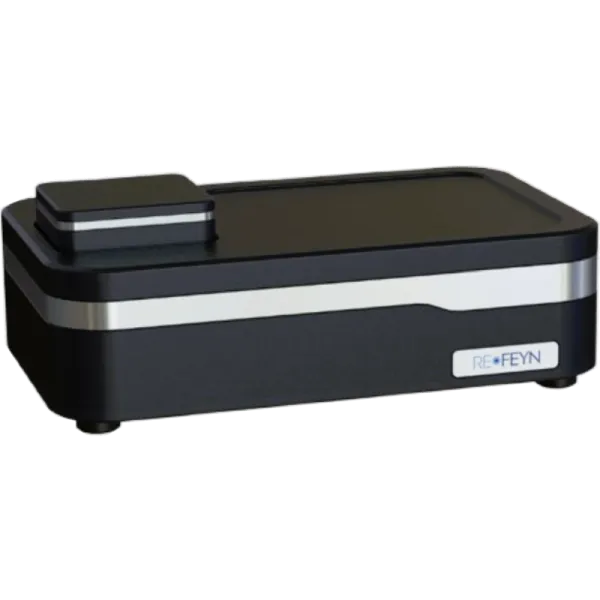
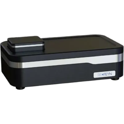
Refeyn Mass Photometer
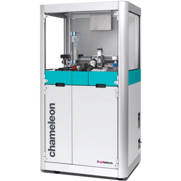
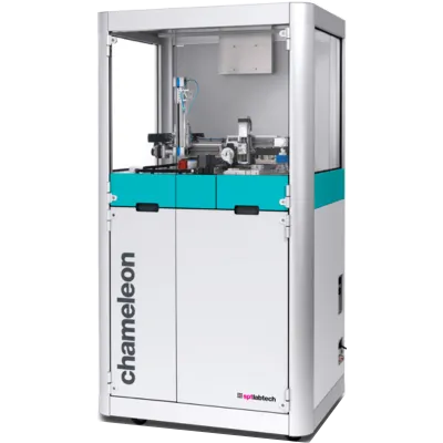
SPT Chameleon
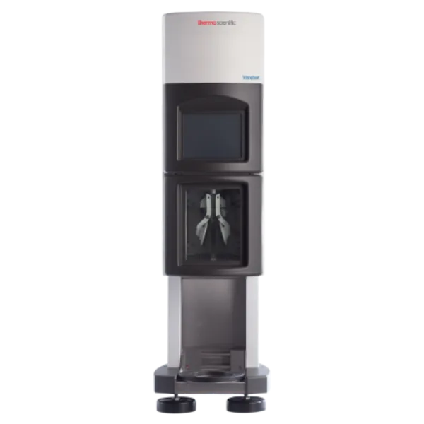
Vitribot
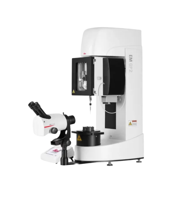
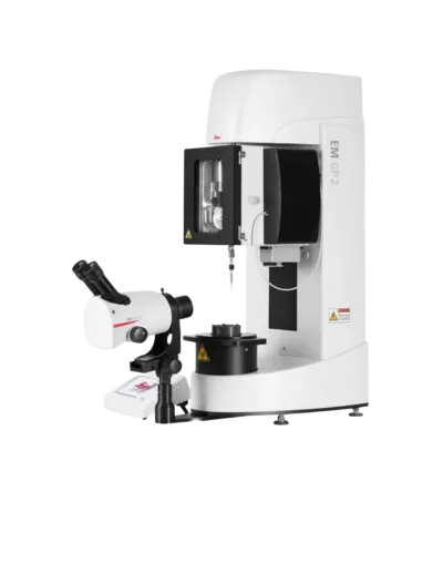
Leica EM GP2
Leica EM GP
Orbitrap Exploris 480
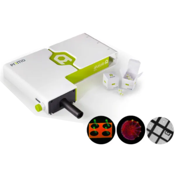
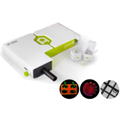
PRIMO micropatterning system
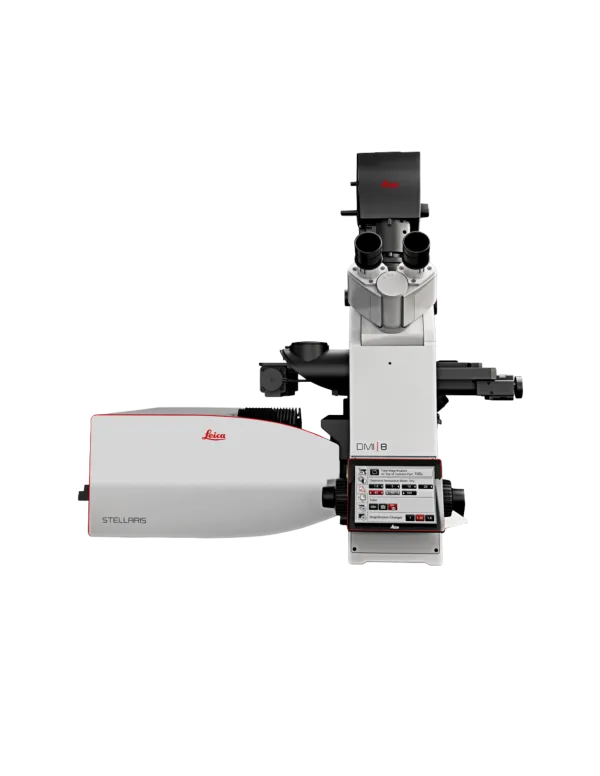
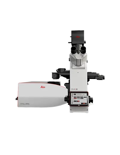
Leica Stellaris 8
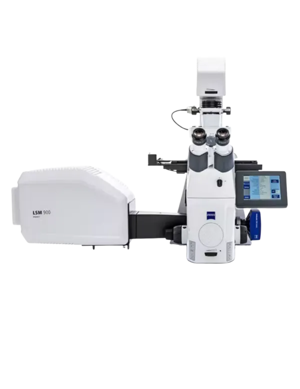
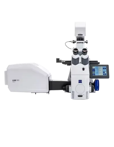
Zeiss LSM 900 with Airyscan 780
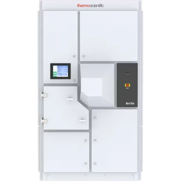
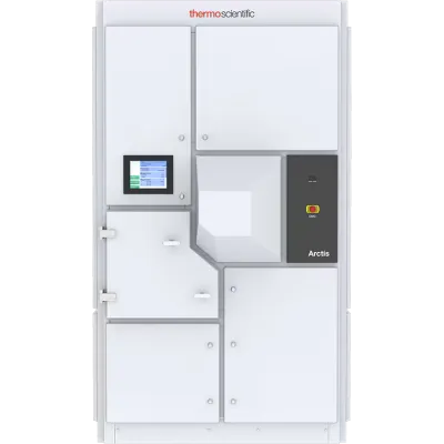
Arctis
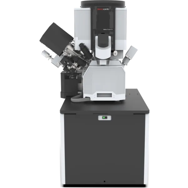
Hydra
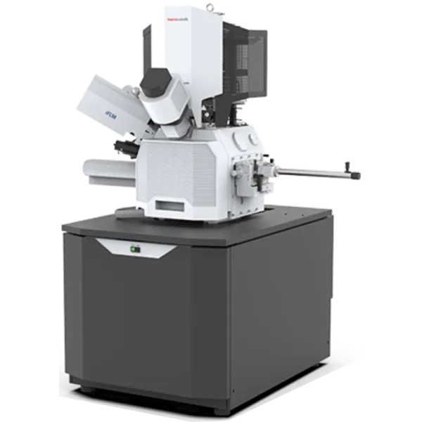
Aquilos2
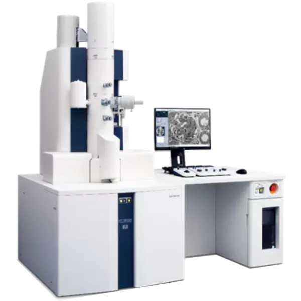
Hitachi 7800
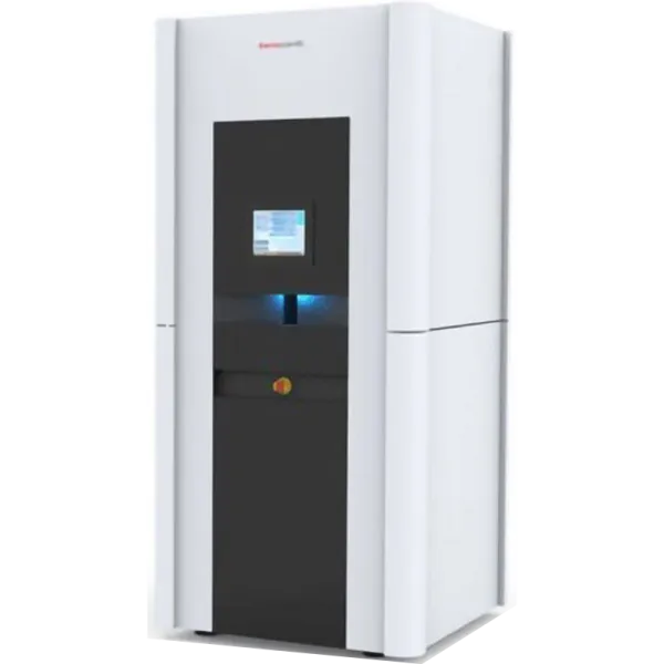
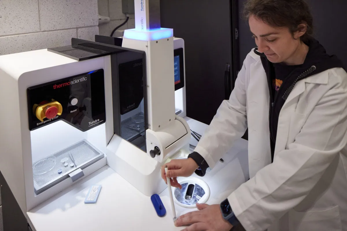
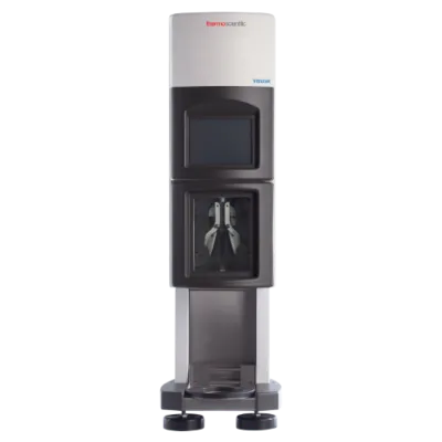
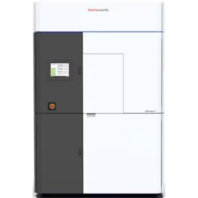
Tundra
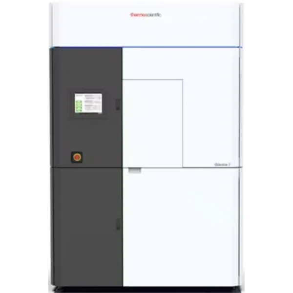
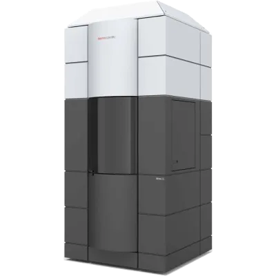
Glacios Electron Microscope
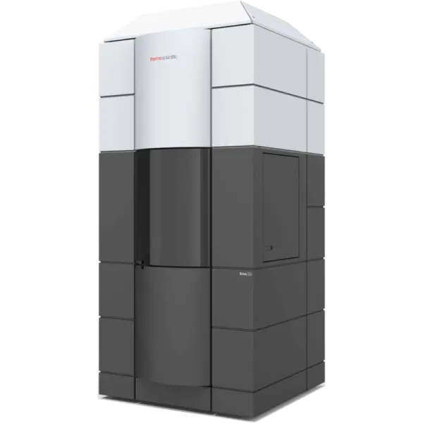
Krios Electron Microscope
Publications
Overcoming air–water interface-induced artifacts in cryo-EM with protein nanocrates
Nature Methods, Vol. 23, Issue 9, Pages 1798-1804
Characterization and modulation of human insulin degrading enzyme conformational dynamics to control enzyme activity
eLife, Vol. 14
Characterization and modulation of human insulin degrading enzyme conformational dynamics to control enzyme activity
eLife, Vol. 14
A bacteriophytochrome Pr/Pfr heterodimer studied through single-particle time-resolved cryo-electron microscopy
Communications Chemistry, Vol. 9, Issue 1
SCM-1/SCAMP Maintains Microdomain Boundaries and Cargo Sorting within the Endosomal System
Simultaneous single-cell profiling of the transcriptome and proteome
Molecular insight into microtubule nucleation by the XMAP215/γ-TuRC module
Nature Communications, Vol. 17, Issue 1
Eg5 activity and density-driven bundling organize the human metaphase mitotic spindle independently of spindle bipolarity
Continuum architecture dynamics of vesicle tethering in exocytosis
Cell, Vol. 189, Issue 4, Pages 1170-1184.e22
A bacteriophytochrome Pr/Pfr heterodimer as obtained by single-ms TR cryo-EM
How augmin establishes the angle of the microtubule branch site
Nature Communications, Vol. 16, Issue 1
Understanding the infection mechanism of Φ8 using infection method in Pseudomonas syringae pv. Phaseolicola strain: In-situ CryoET analysis