
Correlative Light and Electron Microscopy (CLEM)
Access:
Fluorescence-guided targeting for high-resolution cryo-imaging
Correlative light and electron microscopy (CLEM) combines the strengths of both modalities: fluorescence microscopy’s specificity and cellular context with electron microscopy’s structural resolution. Cryo-CLEM extends this to vitrified specimens, eliminating chemical fixation and preserving native cellular organization while enabling precise, light-guided targeting of regions for high-resolution imaging.
NYSBC’s cryo-CLEM facility integrates advanced confocal and super-resolution light microscopy with cryo-EM, enabling scientists to identify structures of interest by fluorescence, then image them at molecular resolution. The approach is particularly powerful for projects where cellular heterogeneity or spatial context is essential, including studies of viral infection, organelle architecture, or protein organization within native environments. NYSBC staff helps researchers develop their proficiency across the full platform and collaborates with them to customize integrated workflows.




User Resources
-
Download the correlative microscopy datasheet for comprehensive technical specifications of SEMC's instrumentation. This resource provides detailed system parameters—including electron energy, Cs values, and detector types—across our Krios and Glacios microscopes, DualBeam FIB-SEM systems, and sample preparation equipment.
-
-
-
Equipment
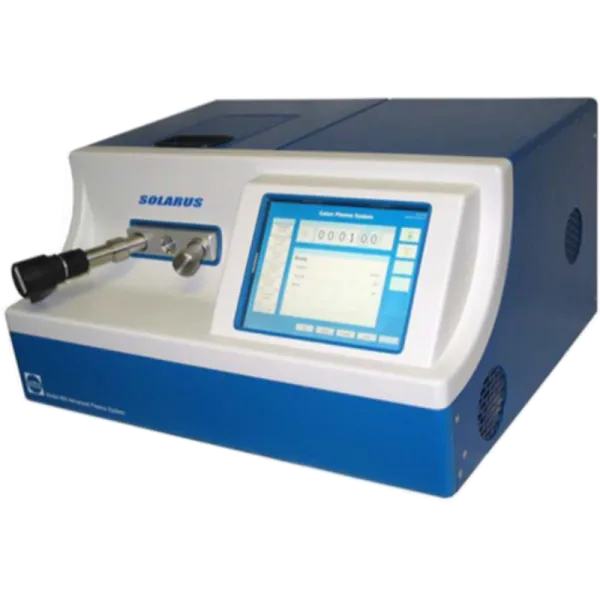
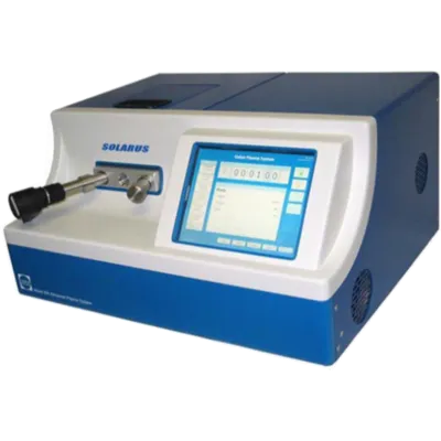
Gatan Solarus
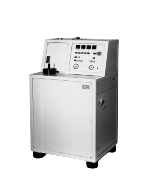
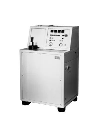
Wohlwend Compac 01
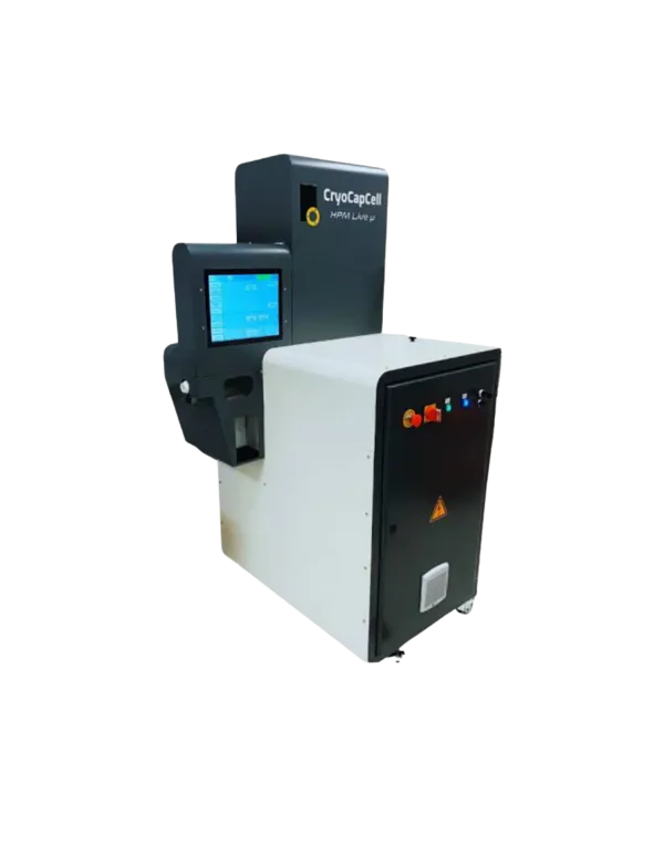
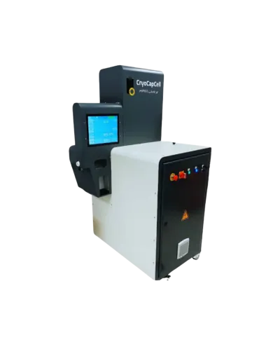
CryoCapCell Live µ
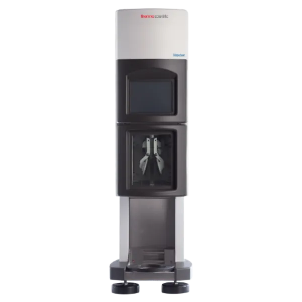
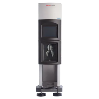
Vitribot
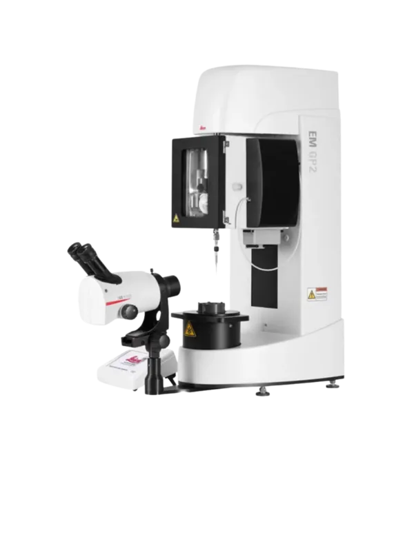
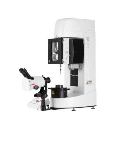
Leica EM GP2
Leica EM GP
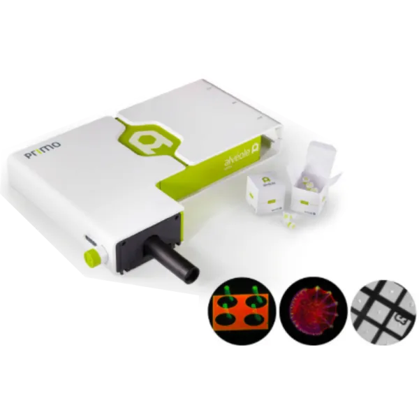
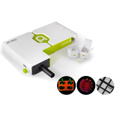
PRIMO micropatterning system
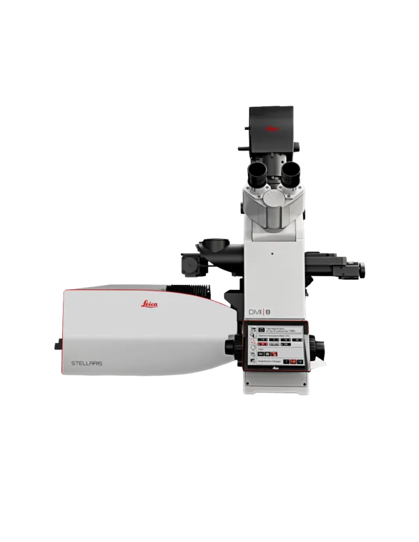
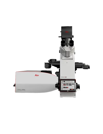
Leica Stellaris 8
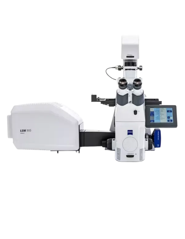
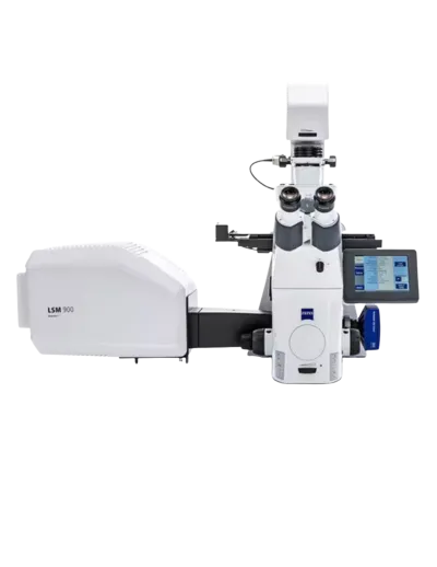
Zeiss LSM 900 with Airyscan 780
Confocal, super-resolution, and cryo-EM on one correlative platform.