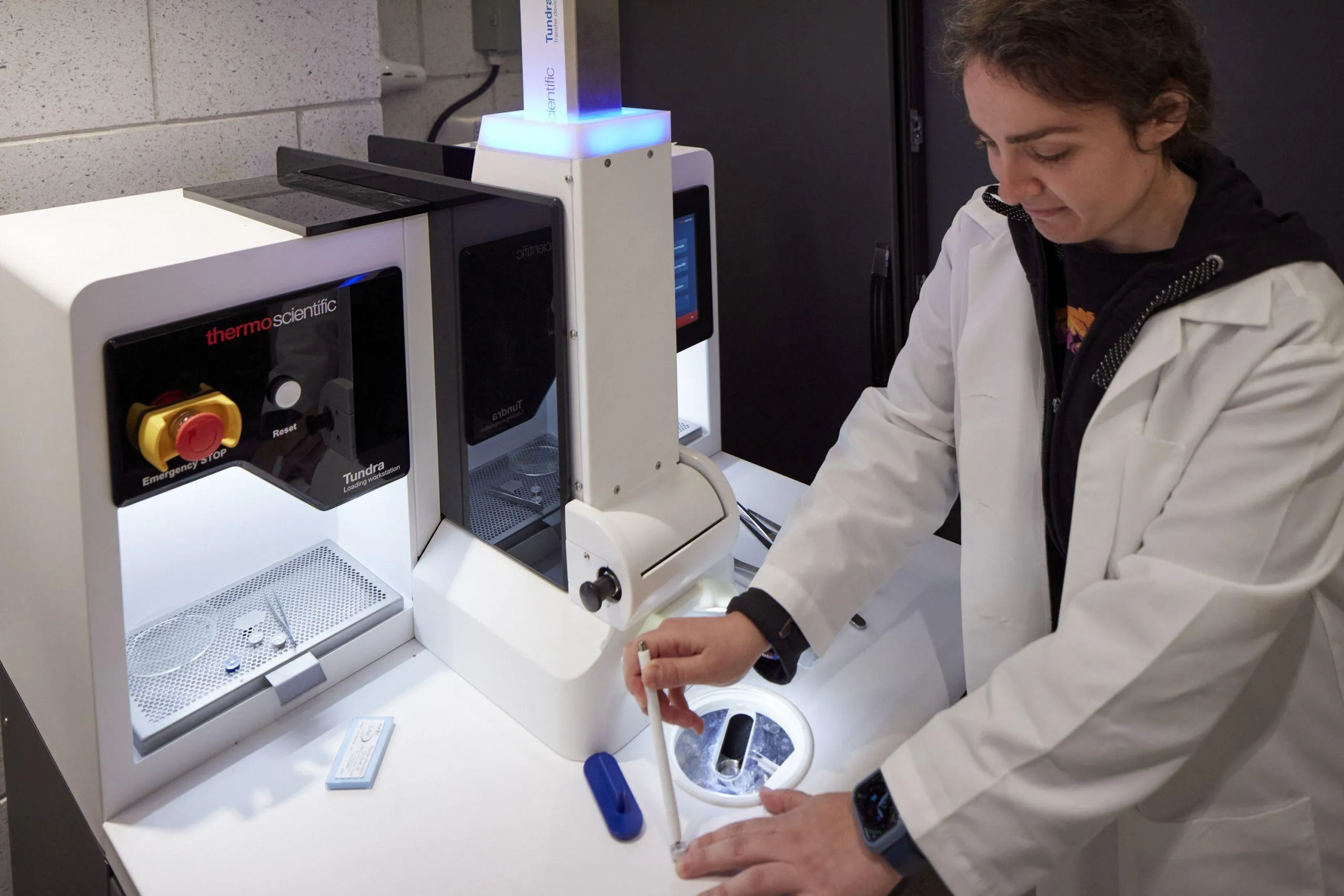
Cryo-Electron Tomography
Access:
Molecular structure in its native cellular context
Cryo-electron tomography (cryo-ET) extends structural biology into the cellular environment, revealing how macromolecular assemblies are organized and function within intact cells and tissues. The technique captures three-dimensional slices through vitrified specimens, preserving native cellular architecture without chemical fixation or heavy-metal staining.
NYSBC supports cryo-ET across two complementary workflows. On-grid tomography works with purified or partially enriched samples, enabling focused structural studies with moderate specimen complexity. In situ approaches—combining cryo-FIB lamella preparation with tomographic imaging—target specific cellular structures directly, from organelles to intact subcellular compartments. Both workflows benefit from integrated sample preparation, correlative light microscopy guidance, and expert staff support.







User Resources
-
Download the cryo-electron tomography datasheet to review the technical specifications for SEMC's tomography-capable instrumentation. This resource outlines system configurations—including specific cryo-ET pixel size calibrations (such as 4Å to 0.8Å on Krios systems), electron energy, and detector types—across our transmission electron microscopes, DualBeam FIB milling systems, and sample preparation equipment.
-
-
-
Equipment
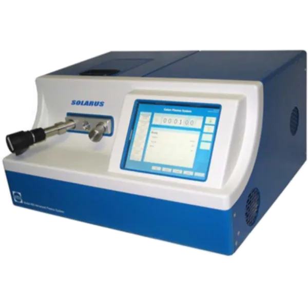
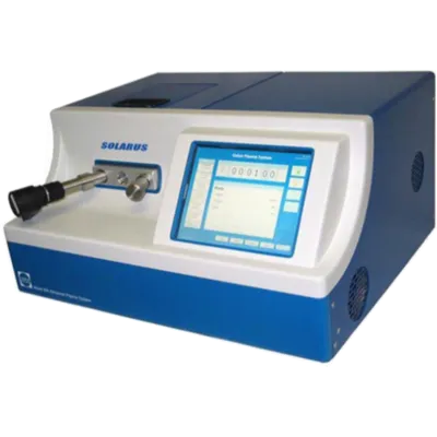
Gatan Solarus
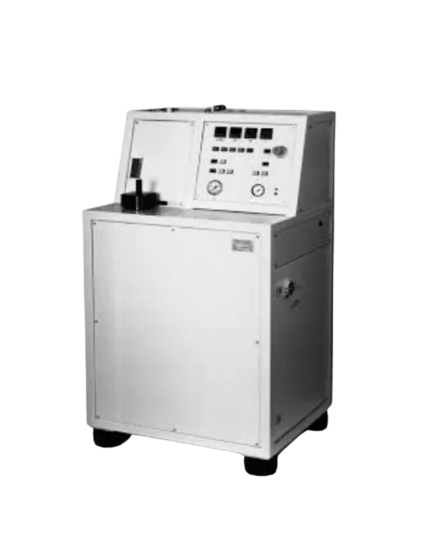
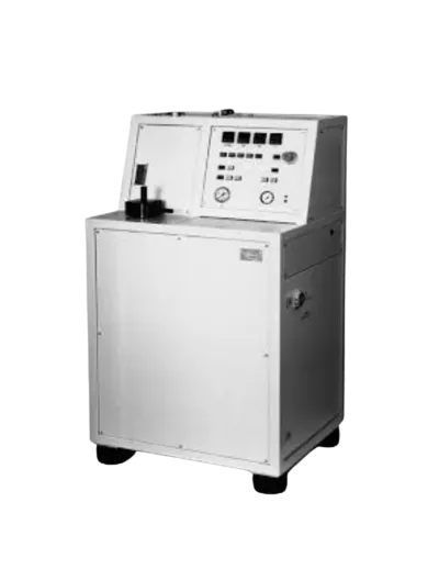
Wohlwend Compac 01
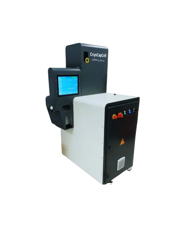
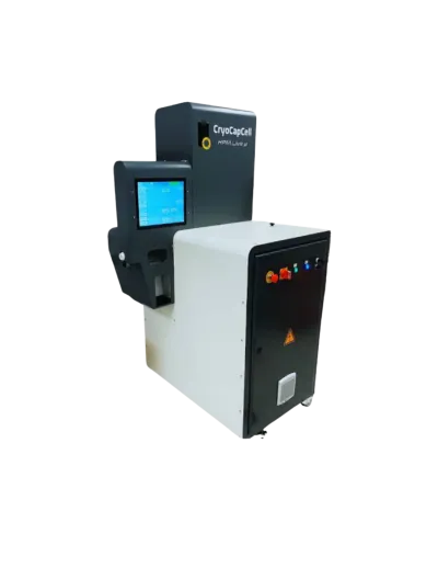
CryoCapCell Live µ
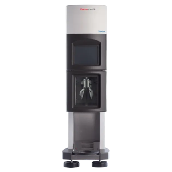
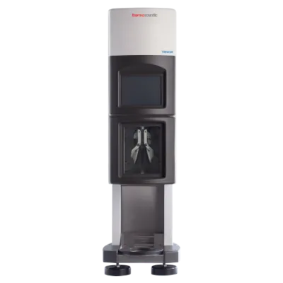
Vitribot
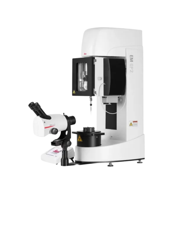
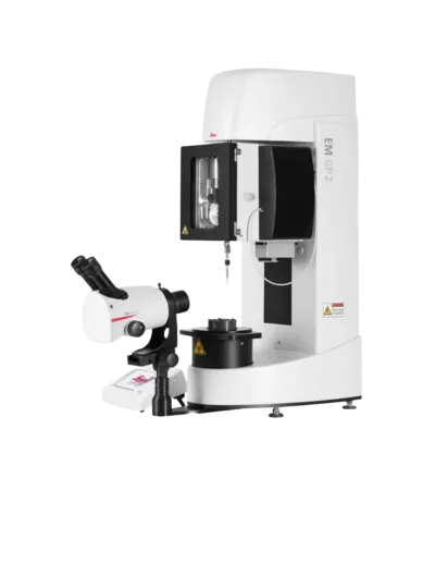
Leica EM GP2
Leica EM GP
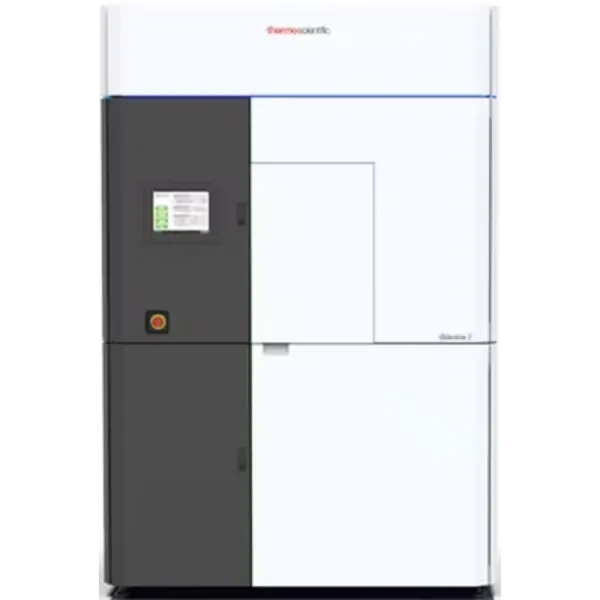
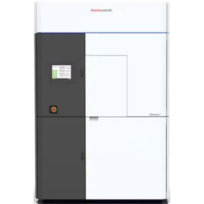
Glacios Electron Microscope
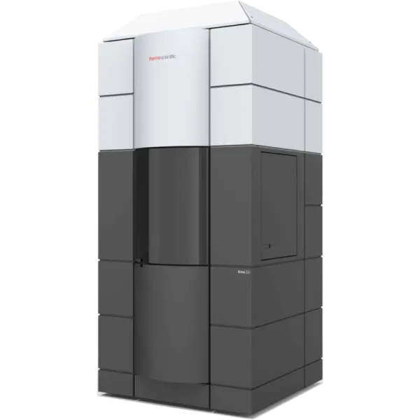
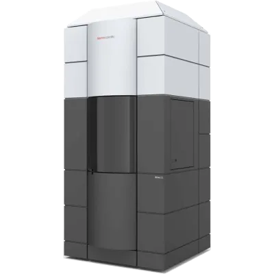
Krios Electron Microscope
“Tomography lets us look at a molecule where it actually works inside the cell, with no fixation and no stain. For a lot of biology, that native context is the whole experiment.”