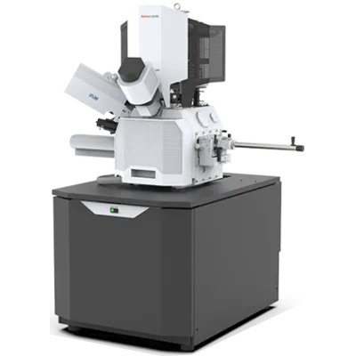
Cryo-FIB (Focused Ion Beam)
Access:
Accessing the cell interior for structural imaging
Cryo-focused ion beam (cryo-FIB) milling enables researchers to prepare thin, electron-transparent lamellae directly from whole cells, tissues, or larger specimens while preserving native structure. By removing excess material while keeping the region of interest intact, cryo-FIB transforms thick biological samples into tractable imaging experiments for high-resolution tomography.
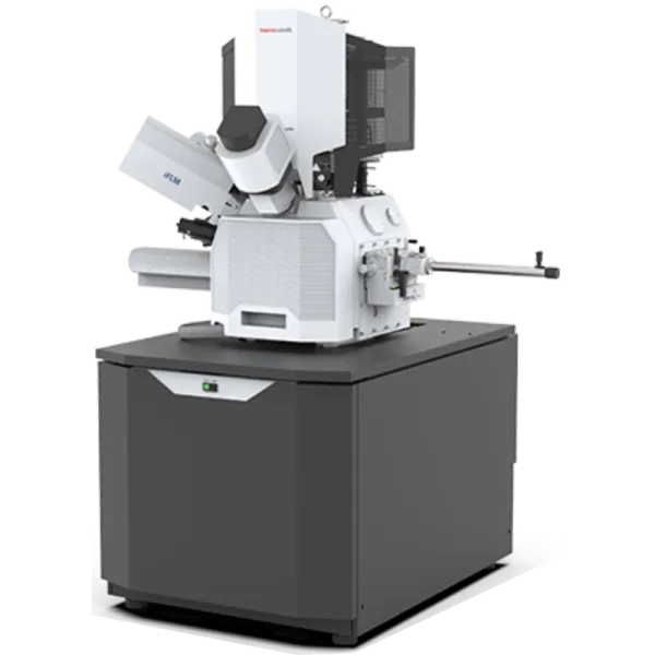
NYSBC operates complementary FIB instruments: the Aquilos2 for high-precision conventional FIB milling and the Hydra and Arctis plasma-FIB systems for faster throughput and advanced workflows. Both platforms support iterative workflows that pair light microscopy targeting with cryo-FIB milling, enabling researchers to locate specific cellular features and prepare them for tomographic imaging. Staff-supported service, hands-on training, and collaborative projects are all available as needed.





User Resources
-
Download the cryo-FIB datasheet for complete technical specifications of SEMC’s focused ion beam milling instrumentation. This document details the configurations of our DualBeam systems—including the Aquilos 2, Hydra, and Arctis—highlighting FIB sources (LMIS and Plasma) and integrated fluorescence light microscopy (iFLM) detectors used for precise lamella preparation.
-
-
-
Equipment
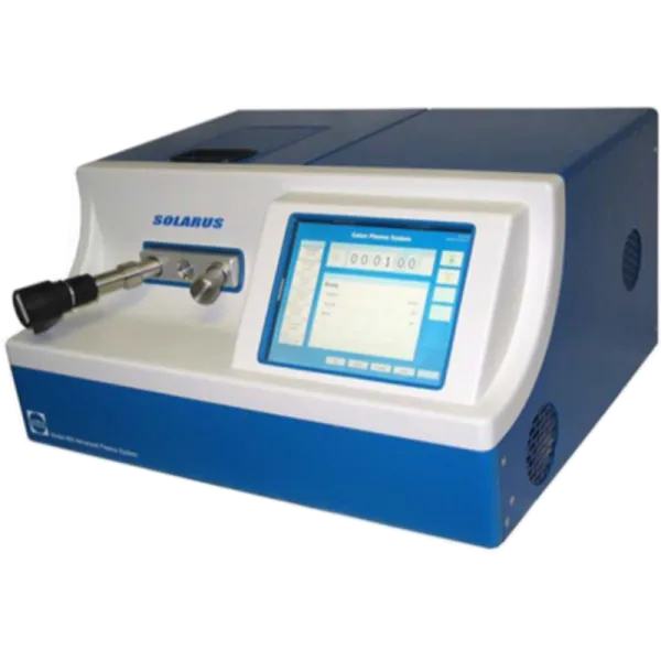
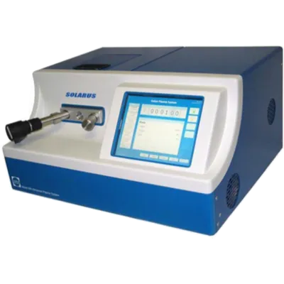
Gatan Solarus
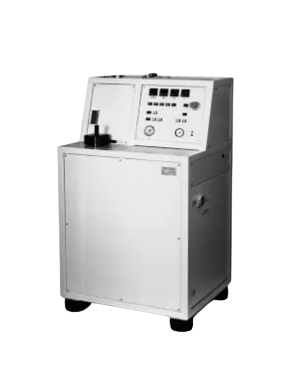
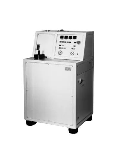
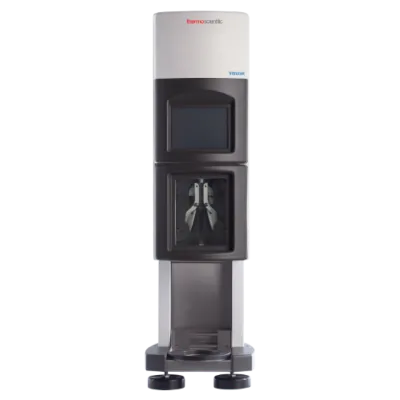
Wohlwend Compac 01
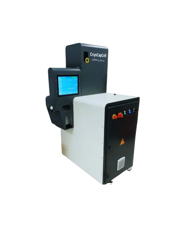
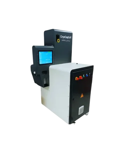
CryoCapCell Live µ
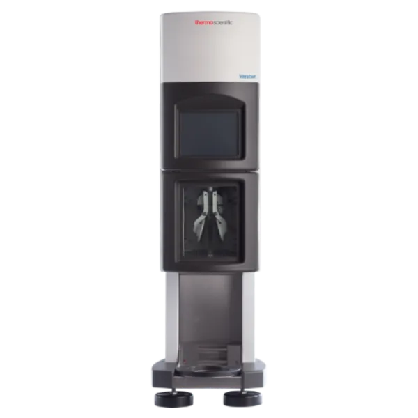
Vitribot
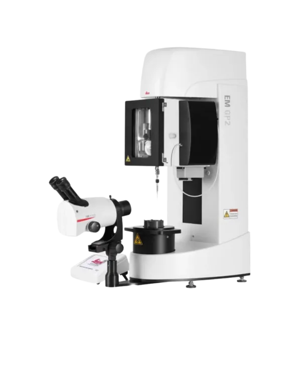
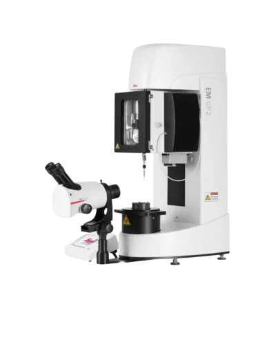
Leica EM GP2
Leica EM GP
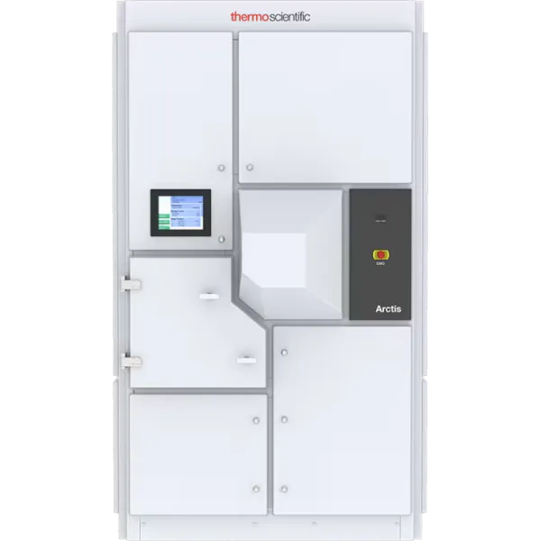
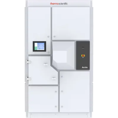
Arctis
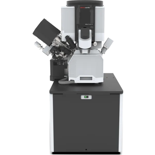
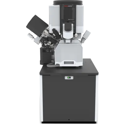
Hydra